These are called varicose veins and they can cause pain and major health issues. The mitral valve is between the left atrium and left ventricle. The aortic semilunar valve prevents backflow from the aorta into the left ventricle. If thechordae tendinae of the mitral valve were damaged, the valve would not function properly and blood could possibly flow backwards towards the pulmonary veins and into the lungs. Answer: The veins from all over the body carry deoxygenated blood to the heart. which specific portion of the question an image, a link, the text, etc your complaint refers to; Varsity Tutors. The impulse spreads through the walls of the right and left atria, causing them to contract, forcing blood into the ventricles. Its the muscle at the centre of your circulation system pumping blood around your body as your heart beats. It prevents the backflow of blood to the right atrium when the right ventricle pumps blood to the lungs.
The pulmonary valve is between the right ventricle and the pulmonary artery. To understand the anatomy and function of the heart, we have divided the heart into two sections - Exterior and Interior. CVI most commonly occurs as the result of a blood clot in the deep veins of the legs a disease known as deep vein thrombosis (DVT). What has valves to prevent backflow blood is at low pressure? Which valves prevent the backflow of blood into the ventricles quizlet? (Arteries dont require valves because pressure from the heart is so strong that blood is only able to flow in one direction.) Other arrhythmias can be serious or even life threatening, such as ventricular arrhythmias. The vena cava is a large vein that brings deoxygenated (impure) blood back to the heart and empties it in to the right atriuma. After picking up oxygen the blood travels back to the heart through the pulmonary veins into the left atrium to the left ventricle and out to the bodys tissues through the aorta. The aortic valve prevents flow from the aorta to the left ventricle and the pulmonary valve prevents flow from the pulmonary artery to the right ventricle. As a result, blood may leak from the ventricle back into the atrium. The adult heart weighs between 200 to 425 grams (7 to 15 ounces) and is about the size of your fist. If you have significant backflow and symptoms, your doctor may prescribe: Surgery is done only if the mitral valve is very abnormal and blood is flowing back into the atrium. They don't have any symptoms or major mitral valve backflow. In fact, most people who have MVP dont have backflow and never have any related symptoms or problems. 101 S. Hanley Rd, Suite 300 misrepresent that a product or activity is infringing your copyrights. Do compression stockings help venous insufficiency? All content 2022Cooper University Health Care. Allegheny College, Bachelor of Science, Neuroscience. After passing through this valve, blood will be in the right ventricle. Your name, address, telephone number and email address; and Veins contain a series of one-way valves and they are squeezed blood is pushed through the valves which then close to prevent backflow. The left atrium collects the oxygenated blood from the lungs, via the pulmonary veins and delivers it to the left ventricle. Blood can even back up from the atrium into the lungs, causing shortness of breath. The tricuspid valve is located between the ventricle and the right atrium.
What prevents backflow of blood inside the heart during contration ? 11. At Cardiac Partners, expert heart surgeons perform mitral valve repair and replacement using minimally invasive techniques. Copyright 2022, All Rights Reserved | Email us: What Prevents The Backflow Of Blood In Veins. The heartbeat is a two part pumping action- Systole (contraction) and Diastole (relaxation). If the heart beats at 70 beats per minute, which of the following is true? On the right side of the heart is the tricuspid valve, composed of three flaps of tissue; on the left is the two-piece mitral valve. Aortic valve: Allows blood to pass from the left ventricle to the aorta; prevents backflow of blood into the left ventricle. Do capillaries have valves to prevent backflow? Physician Referrals and Appointments: If the flow of blood reverses, the flaps fill and are pressed against each other, thus blocking the reentry of blood into the aorta. Blood is supplied to the heart by the coronary arteries. The atrioventricular septum is the muscular wall that divides the right and left sides of the heart. Where is the mitral valve, and what is its function? There are two ventricles, right and left, which are the two lower chambers of the four muscular chambers of the heart.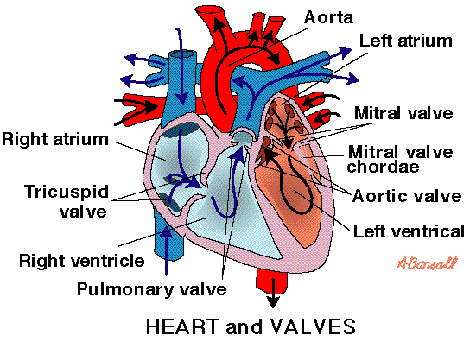 As the heart pumps blood a series of valves open and close tightly. Which blood vessel carries blood for oxidation? The impulse then reaches the atrioventricular (AV) node, which acts as an electrical bridge for impulses to travel from the atria to the ventricles. When the SA node stimulates the heart, it initiated atrial systole. The backflow of blood strains the muscles of both the atrium and the ventricle. In addition to poor cosmesis CVI can lead. As the heart pumps blood, a series of valves open and close tightly. 4. Carries deoxygenated blood from the heart to the lungs. pulmonary vein: One of four veins that carry oxygen-rich blood from the lungs to the heart. The heart acts a pump, delivering blood to the organs, tissues, and cells of your body through a complex network of arteries, arterioles, and capillaries. This causes blood to leak back into the chambers instead of flowing through the heart or into an artery. The right atrium contracts to do this. In people who have MVP, the mitral valve may be abnormal in the following ways: These problems can keep the valve from making a tight seal. There is only one atrial pacemaker region, which ensures coordinated contraction. What is present in veins but absent in arteries? In an average adult, the blood volume is around five liters. Serious heart damage may occur when the coronary circulation is blocked. Right ventricle: Receives blood from the right atrium; pumps blood into the pulmonary artery. The left ventricle collects the pure blood from the left atrium and delivers it to the aorta (main artery) from where it is pumped to the rest of the body. link to the specific question (not just the name of the question) that contains the content and a description of Blood might flow back through thetricuspidvalve to the lungs through the left pulmonary artery. The tricuspid valve prevents backflow from the right ventricle into the right atrium. The valves between the atria and ventricles are called atrioventricular valves (also called cuspid valves) while those at the bases of the large vessels leaving the ventricles are called semilunar valves. If the heart rate is too slow, too fast, or irregular, the heart may not be able to pump enough blood to the body. The P wave of the electrocardiogram corresponds with atrial contraction (atrial systole). In turn, veins bring nutrient-depleted blood back to the heart.
  Valves are present only in the veins and not in, why are swamps more productive than streams. Please be advised that you will be liable for damages (including costs and attorneys fees) if you materially Blood flows from your right atrium into your right ventricle through the. Regurgitation or backflow occurs when the valve does not close tightly.
 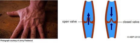 Situated on the wall of the right atrium, this small cluster of specialized cells is the heart's natural pacemaker, initiating electrical impulses at a normal rate. The bicuspid valve prevents backflow from the left ventricle into the left atrium. As a result, the atria aren't able to pump blood into the ventricles the way they should. The valves in the venous system are of this same type. Most people who have the condition are born with it. Rarely, blood can leak the wrong way through the floppy valve. Vasodilators to widen your blood vessels and reduce your hearts workload.
Fortunately therapeutic compression socks have been proven to improve blood circulation and the symptoms of Venous Insufficiency. The deoxygenated blood from the heart muscle is collected by the coronary veins and drained into the right atrium. Corrections? 2. From the right atrium it is pumped to the right ventricle and then to the pulmonary arteries, which carry it to the lungs for reoxygenation. There is a specialized group of cardiac cells responsible for initiating this action potential throughout the heart. They tend to be mild but can worsen over time, mainly when complications occur. Then, the ventricles contract to pump the blood out of the heart. Which valves prevent the backflow of blood to the right ventricle? Which type of blood vessels carry blood away from the heart? improve our educational resources.
Pulmonary arteries: Carry oxygen-depleted blood from the heart to the lungs. Carries deoxygenated blood from the body back to the heart. Blood comes into the right atrium from the body moves into the right ventricle and is pushed into the pulmonary arteries in the lungs. Although the left and right ventricles have chamber volumes of around 100 milliliters, the amount emptied during each heartbeat is only 70% of ventricular volume.
Fortunately. MVP doesnt always cause backflow. The series of activities in systole which happens at one particular moment are: In a normal resting adult, the heart beats about 72 times per minute (Pulse 72), which means all the above activities happen in less than one second. The upper chamber is called the left atrium. What prevents the backflow of blood in the veins class 10? or more of your copyrights, please notify us by providing a written notice (Infringement Notice) containing MVP is the most common heart condition that puts people at risk for this infection. The valve flaps may be too large and thick. The semilunar valves prevent backflow into the ventricles from the aorta and pulmonary arteries. (Palpitations are feelings that your heart is skipping a beat, fluttering, or beating too hard or too fast.). 13. Blood might flow back through the tricuspidvalve to the lungs through the right pulmonary artery. Which blood vessels carry blood for oxidation in Brainly? Gum infections and tooth decay can cause IE. When present, they're most often caused by the backflow of blood through the mitral valve. Carries oxygenated blood from the lungs to the heart. It flows through the right side of the heart, to the lungs, and back to the left side of the heart. The right side of the heart collects oxygen-depleted blood and pumps it to the lungs, through the pulmonary arteries, so that the lungs can refresh the blood with a fresh supply of oxygen. 5. If you have MVP, you can take steps to prevent IE. Mitral valve backflow is most common among men and people who havehigh blood pressure. So it will not be affected by thechordae tendinae of the mitral valve and in any case blood flow in this part of the heart could not flow back to the pulmonary veins. Blood is returned to your heart through venules (small veins) and veins. human cardiovascular system: Valves of the heart, To prevent backflow of blood, the heart is equipped with, https://www.britannica.com/science/valve-anatomy, University of Minnesota - Atlas of Human Cardiac Anatomy - Cardiac Valve Nomenclature. The main function of the heart valves is to regulate and prevent the backflow of the blood. These valves ensure that blood flows in only one direction, preventing backflow. The mitral valve closes and the left ventricle contracts, pumping blood through the aortic valve into the aorta, through which oxygen-richblood is transferred to the whole body. The most common types of arrhythmias are harmless. Which of the followingwould happen if the chordae tendinae attached to the mitral valve were torn or damaged? The heart also has a right atrium and ventricle, separated by the tricuspid valve. From there, it is pumped into the left ventricle and out to the body through the aorta. The atrioventricular (AV) node, bundle of His, and Purkinje fibers are progressively lower in the conduction system and are not associated with P wave generation. your copyright is not authorized by law, or by the copyright owner or such owners agent; (b) that all of the There are two atria, the right atrium, and the left atrium, which are the two upper chambers of the four muscular chambers of the heart. an Pulmonary veins: Deliver oxygen-rich blood from the lungs to the left atrium of the heart. Superior vena cava: Receives blood from the upper body; delivers blood into the right atrium. It is located in the right atrium and generated cardiac action potentials. Which veins valves prevent the backflow of blood and infection? The flaps of the valve are floppy and may not close tightly. Valves are present only in the veins and not in the capillaries and arteries. Then, the tricuspid valve closes and the right ventricle contracts to pump the blood through the pulmonary valve into the pulmonary arteries, which carry oxygen-poorblood into the lungs to be oxygenated. A valve may consist of a sphincter muscle or two or three membranous flaps or folds. Oxygen-rich blood goes to the lungs to be deoxygenated, then returns to the heart, Oxygen-poor blood goes to the lungs to be oxygenated, then returns to the heart, Oxygen-poor blood goes to the heart to be oxygenated, then returns to the body, Oxygen-rich blood goes to the heart to be deoxygenated, then returns to the body. Blood clots can occur because some blood "pools" in the atria instead of flowing into the ventricles. The main function of the pericardium is to: The coronary circulation consists of the blood vessels that supply blood to, and remove blood from, the heart tissue. The myocardium is the layer of the heart that contains the muscle cells. Pulse or Heart Rate is the number of heartbeats per minute. What prevents backflow in veins These are what fail in varicose veins )? IE doesn't happen often, but when it does, it's serious. The cardiac cycle consists of the fillingof the right atrium with venous blood(oxygen-poor blood that has returnedfrom the body to now be pumped into the lungs for oxygenation), and opening of the tricuspid valve to allow transfer of blood to the the right ventricle. AV valves prevent backflow from the ventricles into the atria and semilunar valves prevent backflow from the aortic and pulmonary trunks into the ventricles. People who have MVP and troublesome mitral valve backflow may be treated with medicines, surgery, or both. 1. Deoxygenated blood enters the right atrium through the superior and inferior vena cavae. Recall that the right side of the heart deals with the oxygen-poor blood returned from the systemic circulation; this same blood is then pumped to the lungs to become oxygen-rich. Please refer to the appropriate style manual or other sources if you have any questions. information described below to the designated agent listed below. The series of activities in diastole which happens at one particular moment are: In a normal resting adult, the heart beats about 72 times per minute (Pulse 72), which means all the above activities happens in less than one second. It prevents the backflow of blood to the left atrium when the left ventricle pumps blood through the aorta to the rest of the body. Is Lipodermatosclerosis life threatening? on or linked-to by the Website infringes your copyright, you should consider first contacting an attorney. The aortic valve sits between the left ventricle and the aorta and prevents backflow of blood into the left ventricle after it contracts. Poor circulation also known as Venous Insufficiency has the potential to cause leg pain swelling and fatigue. The bicuspid, or mitral, valve separates the left atrium and ventricle. Some people will need surgery to repair or replace their mitral valves. The presence of symptoms doesnt always mean that the backflow of blood through the valve is significant. This pacemaker structure is called the sinoatrial node. The tricuspid valve is situated between the right atrium and right ventricle. Click here for information on how we use cookies. Some people's valves are abnormal in more than one way. This delivery is regulated by the aortic valve. They are involved in signal mediation and ventricular systole, which corresponds with the QRS complex. Please follow these steps to file a notice: A physical or electronic signature of the copyright owner or a person authorized to act on their behalf; This can lead topalpitations, shortness of breath, chest pain, and other symptoms. Two main coronary arteries branch off the aorta then branch into several smaller arteries that supply oxygen rich blood to the heart. The heart has two types of valves that keep the blood flowing in the correct direction. what impact can the bottleneck effect have on populations.
The heart has a total of four chambers: right atrium, right ventricle, left atrium and left ventricle. From there it leaves the heart via the pulmonary arteries, and enters the pulmonary capillaries. What is the correct route of blood in a human? Also, it's more common in people who are born with connective tissue disorders, such asMarfan syndrome. Mitral valve prolapse (MVP) is a condition in which the hearts mitral valve doesnt work well. Infringement Notice, it will make a good faith attempt to contact the party that made such content available by Which structure is referred to as the pacemaker of the heart? The tricuspid valve prevents backflow of blood from the __________ into the __________. The mitral valve, after the left atrium, is where oxygenated blood arrives when it travels back to the heart from the left and right pulmonary veins. The mitral valve controls blood flow between the upper and lower chambers of the left side of the heart. The aortic valve is between the left ventricle and the aorta. information contained in your Infringement Notice is accurate, and (c) under penalty of perjury, that you are The cardiac sphincter divides the esophagus from the stomach, and is actually part of the digestive system. At a heart rate of 70 beats per minute, then approximately 5 liters is pumped by EACH side of the heart each minute. 3. It pumps blood to all parts of the body through a network of blood vessels by continuously expanding and contracting. They form a tight seal that prevents blood from flowing back into the atria. In AF, the walls of the atria quiver instead of beating normally. Once blood has left the heart and entered the aorta, its return is prevented by the semilunar valves, which consist of membranous saclike flaps that open away from the heart. 7. Valves also help blood travel back to the heart against the force of gravity. Along the way, blood is routed through the kidneys and liver, as well, filtering waste products from the blood. These valves close at the end of systole preventing the backflow of blood from arteries to ventricles and producing the second heart sound. 9. CVI also results from pelvic tumors and vascular malformations and sometimes occurs for unknown reasons.
The pulmonary valve is between the right ventricle and the pulmonary artery. To understand the anatomy and function of the heart, we have divided the heart into two sections - Exterior and Interior. CVI most commonly occurs as the result of a blood clot in the deep veins of the legs a disease known as deep vein thrombosis (DVT). What has valves to prevent backflow blood is at low pressure? Which valves prevent the backflow of blood into the ventricles quizlet? (Arteries dont require valves because pressure from the heart is so strong that blood is only able to flow in one direction.) Other arrhythmias can be serious or even life threatening, such as ventricular arrhythmias. The vena cava is a large vein that brings deoxygenated (impure) blood back to the heart and empties it in to the right atriuma. After picking up oxygen the blood travels back to the heart through the pulmonary veins into the left atrium to the left ventricle and out to the bodys tissues through the aorta. The aortic valve prevents flow from the aorta to the left ventricle and the pulmonary valve prevents flow from the pulmonary artery to the right ventricle. As a result, blood may leak from the ventricle back into the atrium. The adult heart weighs between 200 to 425 grams (7 to 15 ounces) and is about the size of your fist. If you have significant backflow and symptoms, your doctor may prescribe: Surgery is done only if the mitral valve is very abnormal and blood is flowing back into the atrium. They don't have any symptoms or major mitral valve backflow. In fact, most people who have MVP dont have backflow and never have any related symptoms or problems. 101 S. Hanley Rd, Suite 300 misrepresent that a product or activity is infringing your copyrights. Do compression stockings help venous insufficiency? All content 2022Cooper University Health Care. Allegheny College, Bachelor of Science, Neuroscience. After passing through this valve, blood will be in the right ventricle. Your name, address, telephone number and email address; and Veins contain a series of one-way valves and they are squeezed blood is pushed through the valves which then close to prevent backflow. The left atrium collects the oxygenated blood from the lungs, via the pulmonary veins and delivers it to the left ventricle. Blood can even back up from the atrium into the lungs, causing shortness of breath. The tricuspid valve is located between the ventricle and the right atrium.
What prevents backflow of blood inside the heart during contration ? 11. At Cardiac Partners, expert heart surgeons perform mitral valve repair and replacement using minimally invasive techniques. Copyright 2022, All Rights Reserved | Email us: What Prevents The Backflow Of Blood In Veins. The heartbeat is a two part pumping action- Systole (contraction) and Diastole (relaxation). If the heart beats at 70 beats per minute, which of the following is true? On the right side of the heart is the tricuspid valve, composed of three flaps of tissue; on the left is the two-piece mitral valve. Aortic valve: Allows blood to pass from the left ventricle to the aorta; prevents backflow of blood into the left ventricle. Do capillaries have valves to prevent backflow? Physician Referrals and Appointments: If the flow of blood reverses, the flaps fill and are pressed against each other, thus blocking the reentry of blood into the aorta. Blood is supplied to the heart by the coronary arteries. The atrioventricular septum is the muscular wall that divides the right and left sides of the heart. Where is the mitral valve, and what is its function? There are two ventricles, right and left, which are the two lower chambers of the four muscular chambers of the heart.
As the heart pumps blood a series of valves open and close tightly. Which blood vessel carries blood for oxidation? The impulse then reaches the atrioventricular (AV) node, which acts as an electrical bridge for impulses to travel from the atria to the ventricles. When the SA node stimulates the heart, it initiated atrial systole. The backflow of blood strains the muscles of both the atrium and the ventricle. In addition to poor cosmesis CVI can lead. As the heart pumps blood, a series of valves open and close tightly. 4. Carries deoxygenated blood from the heart to the lungs. pulmonary vein: One of four veins that carry oxygen-rich blood from the lungs to the heart. The heart acts a pump, delivering blood to the organs, tissues, and cells of your body through a complex network of arteries, arterioles, and capillaries. This causes blood to leak back into the chambers instead of flowing through the heart or into an artery. The right atrium contracts to do this. In people who have MVP, the mitral valve may be abnormal in the following ways: These problems can keep the valve from making a tight seal. There is only one atrial pacemaker region, which ensures coordinated contraction. What is present in veins but absent in arteries? In an average adult, the blood volume is around five liters. Serious heart damage may occur when the coronary circulation is blocked. Right ventricle: Receives blood from the right atrium; pumps blood into the pulmonary artery. The left ventricle collects the pure blood from the left atrium and delivers it to the aorta (main artery) from where it is pumped to the rest of the body. link to the specific question (not just the name of the question) that contains the content and a description of Blood might flow back through thetricuspidvalve to the lungs through the left pulmonary artery. The tricuspid valve prevents backflow from the right ventricle into the right atrium. The valves between the atria and ventricles are called atrioventricular valves (also called cuspid valves) while those at the bases of the large vessels leaving the ventricles are called semilunar valves. If the heart rate is too slow, too fast, or irregular, the heart may not be able to pump enough blood to the body. The P wave of the electrocardiogram corresponds with atrial contraction (atrial systole). In turn, veins bring nutrient-depleted blood back to the heart. Valves are present only in the veins and not in, why are swamps more productive than streams. Please be advised that you will be liable for damages (including costs and attorneys fees) if you materially Blood flows from your right atrium into your right ventricle through the. Regurgitation or backflow occurs when the valve does not close tightly. Situated on the wall of the right atrium, this small cluster of specialized cells is the heart's natural pacemaker, initiating electrical impulses at a normal rate. The bicuspid valve prevents backflow from the left ventricle into the left atrium. As a result, the atria aren't able to pump blood into the ventricles the way they should. The valves in the venous system are of this same type. Most people who have the condition are born with it. Rarely, blood can leak the wrong way through the floppy valve. Vasodilators to widen your blood vessels and reduce your hearts workload. Fortunately therapeutic compression socks have been proven to improve blood circulation and the symptoms of Venous Insufficiency. The deoxygenated blood from the heart muscle is collected by the coronary veins and drained into the right atrium. Corrections? 2. From the right atrium it is pumped to the right ventricle and then to the pulmonary arteries, which carry it to the lungs for reoxygenation. There is a specialized group of cardiac cells responsible for initiating this action potential throughout the heart. They tend to be mild but can worsen over time, mainly when complications occur. Then, the ventricles contract to pump the blood out of the heart. Which valves prevent the backflow of blood to the right ventricle? Which type of blood vessels carry blood away from the heart? improve our educational resources.
Pulmonary arteries: Carry oxygen-depleted blood from the heart to the lungs. Carries deoxygenated blood from the body back to the heart. Blood comes into the right atrium from the body moves into the right ventricle and is pushed into the pulmonary arteries in the lungs. Although the left and right ventricles have chamber volumes of around 100 milliliters, the amount emptied during each heartbeat is only 70% of ventricular volume.
Fortunately. MVP doesnt always cause backflow. The series of activities in systole which happens at one particular moment are: In a normal resting adult, the heart beats about 72 times per minute (Pulse 72), which means all the above activities happen in less than one second. The upper chamber is called the left atrium. What prevents the backflow of blood in the veins class 10? or more of your copyrights, please notify us by providing a written notice (Infringement Notice) containing MVP is the most common heart condition that puts people at risk for this infection. The valve flaps may be too large and thick. The semilunar valves prevent backflow into the ventricles from the aorta and pulmonary arteries. (Palpitations are feelings that your heart is skipping a beat, fluttering, or beating too hard or too fast.). 13. Blood might flow back through the tricuspidvalve to the lungs through the right pulmonary artery. Which blood vessels carry blood for oxidation in Brainly? Gum infections and tooth decay can cause IE. When present, they're most often caused by the backflow of blood through the mitral valve. Carries oxygenated blood from the lungs to the heart. It flows through the right side of the heart, to the lungs, and back to the left side of the heart. The right side of the heart collects oxygen-depleted blood and pumps it to the lungs, through the pulmonary arteries, so that the lungs can refresh the blood with a fresh supply of oxygen. 5. If you have MVP, you can take steps to prevent IE. Mitral valve backflow is most common among men and people who havehigh blood pressure. So it will not be affected by thechordae tendinae of the mitral valve and in any case blood flow in this part of the heart could not flow back to the pulmonary veins. Blood is returned to your heart through venules (small veins) and veins. human cardiovascular system: Valves of the heart, To prevent backflow of blood, the heart is equipped with, https://www.britannica.com/science/valve-anatomy, University of Minnesota - Atlas of Human Cardiac Anatomy - Cardiac Valve Nomenclature. The main function of the heart valves is to regulate and prevent the backflow of the blood. These valves ensure that blood flows in only one direction, preventing backflow. The mitral valve closes and the left ventricle contracts, pumping blood through the aortic valve into the aorta, through which oxygen-richblood is transferred to the whole body. The most common types of arrhythmias are harmless. Which of the followingwould happen if the chordae tendinae attached to the mitral valve were torn or damaged? The heart also has a right atrium and ventricle, separated by the tricuspid valve. From there, it is pumped into the left ventricle and out to the body through the aorta. The atrioventricular (AV) node, bundle of His, and Purkinje fibers are progressively lower in the conduction system and are not associated with P wave generation. your copyright is not authorized by law, or by the copyright owner or such owners agent; (b) that all of the There are two atria, the right atrium, and the left atrium, which are the two upper chambers of the four muscular chambers of the heart. an Pulmonary veins: Deliver oxygen-rich blood from the lungs to the left atrium of the heart. Superior vena cava: Receives blood from the upper body; delivers blood into the right atrium. It is located in the right atrium and generated cardiac action potentials. Which veins valves prevent the backflow of blood and infection? The flaps of the valve are floppy and may not close tightly. Valves are present only in the veins and not in the capillaries and arteries. Then, the tricuspid valve closes and the right ventricle contracts to pump the blood through the pulmonary valve into the pulmonary arteries, which carry oxygen-poorblood into the lungs to be oxygenated. A valve may consist of a sphincter muscle or two or three membranous flaps or folds. Oxygen-rich blood goes to the lungs to be deoxygenated, then returns to the heart, Oxygen-poor blood goes to the lungs to be oxygenated, then returns to the heart, Oxygen-poor blood goes to the heart to be oxygenated, then returns to the body, Oxygen-rich blood goes to the heart to be deoxygenated, then returns to the body. Blood clots can occur because some blood "pools" in the atria instead of flowing into the ventricles. The main function of the pericardium is to: The coronary circulation consists of the blood vessels that supply blood to, and remove blood from, the heart tissue. The myocardium is the layer of the heart that contains the muscle cells. Pulse or Heart Rate is the number of heartbeats per minute. What prevents backflow in veins These are what fail in varicose veins )? IE doesn't happen often, but when it does, it's serious. The cardiac cycle consists of the fillingof the right atrium with venous blood(oxygen-poor blood that has returnedfrom the body to now be pumped into the lungs for oxygenation), and opening of the tricuspid valve to allow transfer of blood to the the right ventricle. AV valves prevent backflow from the ventricles into the atria and semilunar valves prevent backflow from the aortic and pulmonary trunks into the ventricles. People who have MVP and troublesome mitral valve backflow may be treated with medicines, surgery, or both. 1. Deoxygenated blood enters the right atrium through the superior and inferior vena cavae. Recall that the right side of the heart deals with the oxygen-poor blood returned from the systemic circulation; this same blood is then pumped to the lungs to become oxygen-rich. Please refer to the appropriate style manual or other sources if you have any questions. information described below to the designated agent listed below. The series of activities in diastole which happens at one particular moment are: In a normal resting adult, the heart beats about 72 times per minute (Pulse 72), which means all the above activities happens in less than one second. It prevents the backflow of blood to the left atrium when the left ventricle pumps blood through the aorta to the rest of the body. Is Lipodermatosclerosis life threatening? on or linked-to by the Website infringes your copyright, you should consider first contacting an attorney. The aortic valve sits between the left ventricle and the aorta and prevents backflow of blood into the left ventricle after it contracts. Poor circulation also known as Venous Insufficiency has the potential to cause leg pain swelling and fatigue. The bicuspid, or mitral, valve separates the left atrium and ventricle. Some people will need surgery to repair or replace their mitral valves. The presence of symptoms doesnt always mean that the backflow of blood through the valve is significant. This pacemaker structure is called the sinoatrial node. The tricuspid valve is situated between the right atrium and right ventricle. Click here for information on how we use cookies. Some people's valves are abnormal in more than one way. This delivery is regulated by the aortic valve. They are involved in signal mediation and ventricular systole, which corresponds with the QRS complex. Please follow these steps to file a notice: A physical or electronic signature of the copyright owner or a person authorized to act on their behalf; This can lead topalpitations, shortness of breath, chest pain, and other symptoms. Two main coronary arteries branch off the aorta then branch into several smaller arteries that supply oxygen rich blood to the heart. The heart has two types of valves that keep the blood flowing in the correct direction. what impact can the bottleneck effect have on populations.
The heart has a total of four chambers: right atrium, right ventricle, left atrium and left ventricle. From there it leaves the heart via the pulmonary arteries, and enters the pulmonary capillaries. What is the correct route of blood in a human? Also, it's more common in people who are born with connective tissue disorders, such asMarfan syndrome. Mitral valve prolapse (MVP) is a condition in which the hearts mitral valve doesnt work well. Infringement Notice, it will make a good faith attempt to contact the party that made such content available by Which structure is referred to as the pacemaker of the heart? The tricuspid valve prevents backflow of blood from the __________ into the __________. The mitral valve, after the left atrium, is where oxygenated blood arrives when it travels back to the heart from the left and right pulmonary veins. The mitral valve controls blood flow between the upper and lower chambers of the left side of the heart. The aortic valve is between the left ventricle and the aorta. information contained in your Infringement Notice is accurate, and (c) under penalty of perjury, that you are The cardiac sphincter divides the esophagus from the stomach, and is actually part of the digestive system. At a heart rate of 70 beats per minute, then approximately 5 liters is pumped by EACH side of the heart each minute. 3. It pumps blood to all parts of the body through a network of blood vessels by continuously expanding and contracting. They form a tight seal that prevents blood from flowing back into the atria. In AF, the walls of the atria quiver instead of beating normally. Once blood has left the heart and entered the aorta, its return is prevented by the semilunar valves, which consist of membranous saclike flaps that open away from the heart. 7. Valves also help blood travel back to the heart against the force of gravity. Along the way, blood is routed through the kidneys and liver, as well, filtering waste products from the blood. These valves close at the end of systole preventing the backflow of blood from arteries to ventricles and producing the second heart sound. 9. CVI also results from pelvic tumors and vascular malformations and sometimes occurs for unknown reasons.